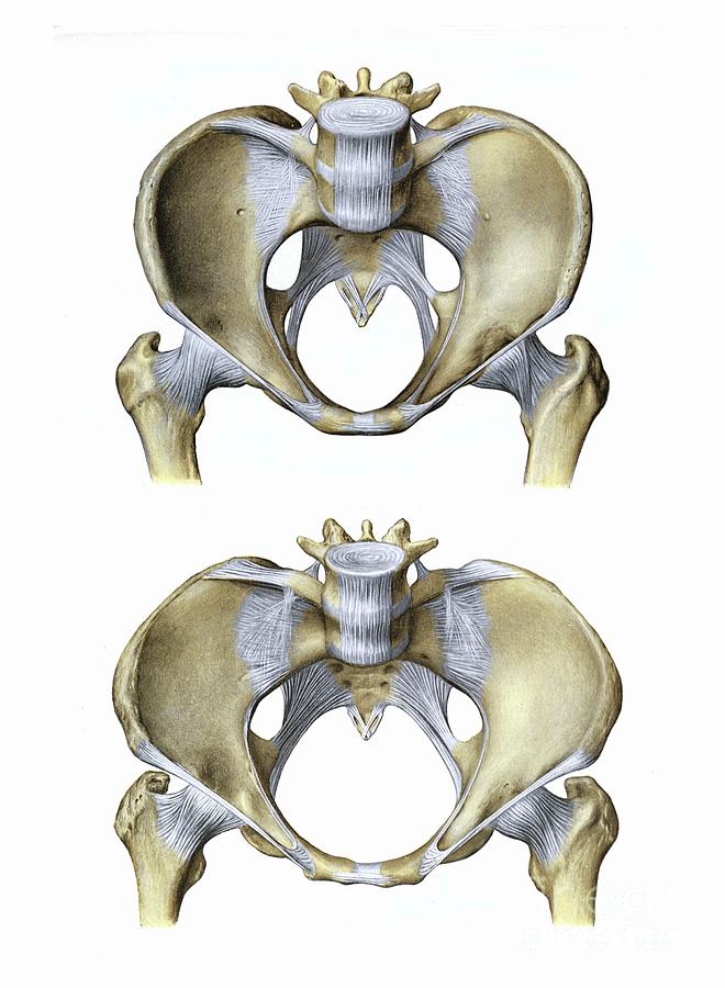
Pelvic Anatomy Ligaments - Ligaments - Posterior Pelvis | Donny Perry | Flickr - Joints and ligaments of the pelvis (anterior view). This image added by admin. The pelvic girdle and pelvic spine. The broad ligament is related to many structures within the female pelvis. The pelvic ligaments are strong, thick bands of fibrous tissue that connect the pelvic bones. The ligaments of the pelvis, are amongst the strongest in the human body.
The broad ligament is a flat sheet of peritoneum, associated with the uterus, fallopian tubes and ovaries. We hope you can get the exact information you. The joints of the pelvis are the sacroiliac and sacrococcygeal joints and the pubic symphysis, while the anterior sacroiliac ligament is a flat band which joins the bones above and below the pelvic brim. The inlet to the pelvic canal is at the level of the sacral promontory and superior aspect of the pubic bones. Pelvic ligaments anatomy instrument cannulating external os of uterus, contrast within uterine cavity, contrast medium in pelvic cavity, contrast within uterine tubes, suspensory ligament of ovary (containing ovarian vessels), infundibulum of uterine tube, posterior lamina of broad ligament, uterine artery and venous plexus, vaginal artery, round ligament, external iliac vessels, fimbriae of.
Unit 26: The Pelvis - Anatomy M1 with Segal at Saint Louis ... from classconnection.s3.amazonaws.com These ligaments are important stabilizers. This image shows the posterior back view of the female pelvic brim (the bones and ligaments that forms the pelvic region in the female) showing: Imaios and selected third parties, use cookies or similar technologies, in particular for audience measurement. We hope you can get the exact information you. This is part of the forced closure method that the pelvis adopts in order to keep itself secure. It extends to both sides of the pelvic wall. There are two major groups of ligaments that provide nearly all the structure of the pelvis. It is close to the major vasculature of the pelvis, including external iliac vein.
The inlet to the pelvic canal is at the level of the sacral promontory and superior aspect of the pubic bones.
This image shows the posterior back view of the female pelvic brim (the bones and ligaments that forms the pelvic region in the female) showing: Cardinal ligament and the uterosacral ligaments provide apical support for the uterus and upper vagina. citation needed this facilitates reconstruction of the floor of the inguinal canal. This image shows the boundaries of the pelvic area formed of the pelvic bones and ligaments showing: The pelvis is the lower portion of the trunk, located between the abdomen and the lower limbs. The outlet is formed by the pubic arch, ischial spines, sacrotuberous ligaments, and the coccyx. Anatomynote.com found pelvis and ligaments cadaver diagram from plenty of anatomical pictures on the internet. Imaios and selected third parties, use cookies or similar technologies, in particular for audience measurement. The cardinal ligament is a paired thickening of the parametrium and pelvic fascia at the base of the broad ligament, which extends between the cervix and vaginal fornix medially to the sidewall of pelvis laterally. The pelvis is held together by three principal ligaments: Cookies allow us to analyze and store information such as the characteristics of your device as well as certain personal data (e.g., ip addresses, navigation, usage or geolocation data, unique identifiers). We think this is the most useful anatomy picture that you need. The femoral ligaments act to stabilize the ball and socket joint of the hip, connecting to the ilium and the ischium.
The pelvis is a boney structure at the base of the lumbar spine. The ilium, ischium and the pubic bone. The pelvis is the lower portion of the trunk, located between the abdomen and the lower limbs. Joints and ligaments of the pelvis the two sacroiliac joints are synovial joints, and are further strengthened by the very strong posterior sacroiliac ligaments which run along the posterior aspect of the joint. There are two major groups of ligaments that provide nearly all the structure of the pelvis.
Pelvic Ligaments Photograph by Microscape/science Photo ... from images.fineartamerica.com Inherent stability of the pelvis is provided by ligaments. Pelvic bone and ligaments anatomy. The cardinal ligaments, also known as the transverse cervical ligaments, the lateral cervical ligaments, or mackenrodt's ligaments, are fibrous bands that attached the cervix to the lateral pelvic walls. It extends from the lateral pelvic walls on both sides, and folds over the internal female genitalia, covering their surface anteriorly and posteriorly. Pelvis anatomy hip anatomy anatomy bones human body anatomy human anatomy and physiology muscle anatomy anatomy study medical anatomy massage therapy. The ligaments of the pelvis, are amongst the strongest in the human body. Bones and ligaments of the female pelvis. Sagittal section through pelvis (gilroy et al.) atlas of anatomy 2nd ed., fig.
Cardinal ligament and the uterosacral ligaments provide apical support for the uterus and upper vagina.
Iliolumbar, sacrotuberous and sacrospinous ligaments. There are two major groups of ligaments that provide nearly all the structure of the pelvis. We think this is the most useful anatomy picture that you need. Joints and ligaments of the pelvis the two sacroiliac joints are synovial joints, and are further strengthened by the very strong posterior sacroiliac ligaments which run along the posterior aspect of the joint. The pelvis consists of two innominate bones and the sacrum to which coccyx is attached. Bones and ligaments of the female pelvis. We are developing an accurate 3d model of human anatomy. The broad ligament can be further divided into three components. The ligaments of the pelvis, are amongst the strongest in the human body. The broad ligament is a flat sheet of peritoneum, associated with the uterus, fallopian tubes and ovaries. The floor of the pelvis is made up of the muscles of the pelvis, which support its. The pelvis's frame is made up of the bones of the pelvis, which connect the axial skeleton to the femurs, and therefore acts in weight bearing of the upper body. In fact, the most important factor stabilizing the pelvic ring structure is the ligaments that hold the two innominate bones and the sacrum together.
The uterosacral ligaments were the most rigid whether at low or high deformation, while the round ligament was more rigid than the broad ligament. Sacrotuberous ligament sacrospinous ligament lesser sciatic foramen greater sciatic foramen medial attachment: Inherent stability of the pelvis is provided by ligaments. Joints and ligaments of the pelvis (anterior view) You can click the image to magnify if you cannot see clearly.
Flashcards - Gross: Male & Female Pelvic Viscera ... from classconnection.s3.amazonaws.com They form what can be described as a basket weave formation, in order to create strength and tensegrity within the structure. Pelvic bone and ligaments anatomy. This image shows the posterior back view of the female pelvic brim (the bones and ligaments that forms the pelvic region in the female) showing: It extends from the lateral pelvic walls on both sides, and folds over the internal female genitalia, covering their surface anteriorly and posteriorly. The pelvis is a boney structure at the base of the lumbar spine. Imaios and selected third parties, use cookies or similar technologies, in particular for audience measurement. This image shows the boundaries of the pelvic area formed of the pelvic bones and ligaments showing: The femoral ligaments act to stabilize the ball and socket joint of the hip, connecting to the ilium and the ischium.
Cardinal ligament and the uterosacral ligaments provide apical support for the uterus and upper vagina.
Pelvis anatomy hip anatomy anatomy bones human body anatomy human anatomy and physiology muscle anatomy anatomy study medical anatomy massage therapy. The pectineal ligament is usually around 6 cm long in adults. The sacral ligaments are responsible for the major connection between the three bones of the pelvis. The pelvis's frame is made up of the bones of the pelvis, which connect the axial skeleton to the femurs, and therefore acts in weight bearing of the upper body. The pubocervical ligaments are a pair of fibrous bands that attach the anterior portion of the cervix to the posterior pubic symphysis. The floor of the pelvis is made up of the muscles of the pelvis, which support its. The pelvis itself is a paired composite structure made up by three bones (ilium, ischium and pubis) that articulates with the sacral part of the axial spine. The inlet to the pelvic canal is at the level of the sacral promontory and superior aspect of the pubic bones. The cardinal ligament is a paired thickening of the parametrium and pelvic fascia at the base of the broad ligament, which extends between the cervix and vaginal fornix medially to the sidewall of pelvis laterally. Additional ligaments may be found in the female pelvis. Pelvic bone and ligaments anatomy. The uterosacral ligament supports the uterus posteriorly, and the pubocervical ligament anchors the uterus anteriorly. This will be explored further on.
They form what can be described as a basket weave formation, in order to create strength and tensegrity within the structure pelvic anatomy. Broad ligament the broad ligament supports the uterus, fallopian tubes, and ovaries.


0 Komentar