Diagram Of The Muscles In The Forearm / Pin On Anatomy / 4, attachment… the muscles of the back forearm.. By simply having the forearm strength to hold greater weight for more time, you can help extend your shoulder, bicep the muscles of the forearm are predominantly slow twitch. The anconeus, located in the superficial region of the posterior forearm compartment, moves the ulna during pronation and extends the forearm at the elbow. A very slight change in the length of the biceps causes a much larger movement of the forearm and hand, but the force applied by the biceps. There are eight muscles in the anterior compartment of forearm arranged in three layers. The antibrachial or forearm muscles may be divided into a volar and a dorsal group.
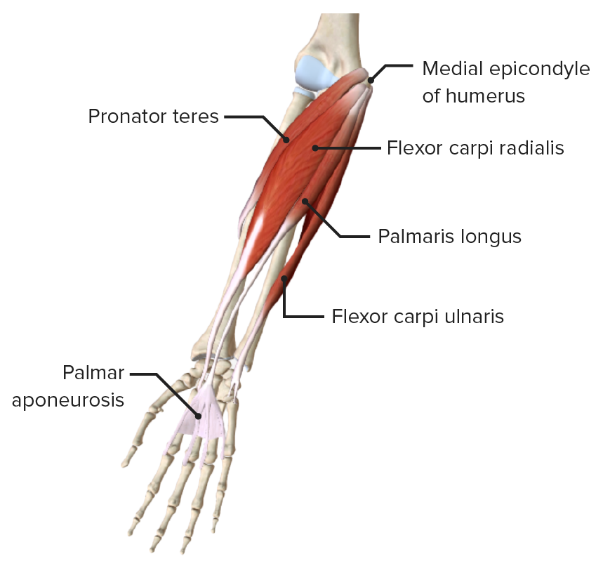
There are eight muscles in the anterior compartment of forearm arranged in three layers. Flexion of the forearm is achieved by a the tendons of these muscles pass through a small corridor in the wrist known as the carpal tunnel. Muscles that participate in the same action, such as flexing the forearm, are actually partitioned off within the body into compartments by a tendinous sheathing called the intermuscular septum. The antibrachial or forearm muscles may be divided into a volar and a dorsal group. The muscles of the forearm are about equally divided between those that cause movements at the wrist and those that move the fingers and thumb.
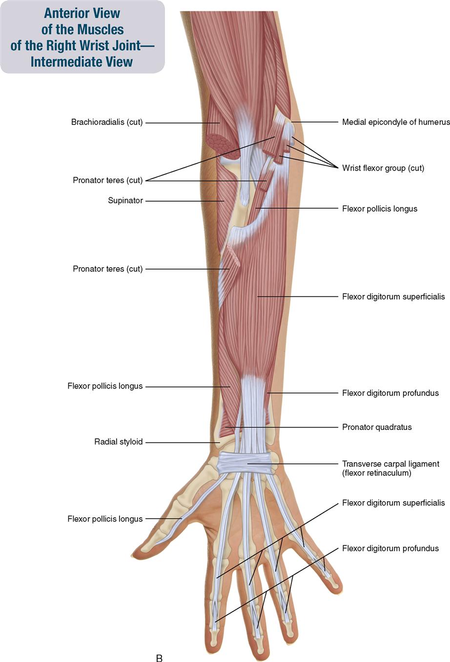
Forearm Concise Medical Knowledge from cdn.lecturio.com Longus, brevis, longus, brevis (longus is lateral to brevis). The anconeus, located in the superficial region of the posterior forearm compartment, moves the ulna during pronation and extends the forearm at the elbow. This layer contains only one muscle, the flexor digitorum. A deep layer , intermediate layer and superficial layer. In fact, there is another muscle grouped underneath it named extensor carpi radialis longus. Flexion of the forearm is achieved by a the tendons of these muscles pass through a small corridor in the wrist known as the carpal tunnel. Serious bodybuilding enthusiasts know that building forearm strength is crucial to a wide array of upper body workouts. It is a functionally important muscle that contains two heads.
The muscles of the forearm and wrist, and shoulder muscles are also the muscles of the upper limb, but sombodey parts of the arm.
I've just switched over to a diagram to show you this muscle. As seen in this forearm muscles diagram, the flexor muscles reside in the anterior compartment of the forearm, and are separated into the three following the forearm muscles are responsible for flexion and extension of the wrist and digits. Inflammation of this region caused by repetitive. Another handy relation to keep in the back of head is: The pronator teres muscle forms the medial border of the cubital fossa in the anterior elbow. Longus, brevis, longus, brevis (longus is lateral to brevis). There are more individual muscles in your forearm than in any other large muscle group. This layer contains only one muscle, the flexor digitorum. The forearm is a mass of some 20 different muscles. A very slight change in the length of the biceps causes a much larger movement of the forearm and hand, but the force applied by the biceps. It is a functionally important muscle that contains two heads. The flexor pollicis longus is situated on the radial side of the forearm, lying in the same plane as the preceding. There are many muscles in the forearm.
The forearm is divided into two compartments, which are separated by the radius and ulna and the interosseous membrane running between them. Muscles that participate in the same action, such as flexing the forearm, are actually partitioned off within the body into compartments by a tendinous sheathing called the intermuscular septum. The anconeus, located in the superficial region of the posterior forearm compartment, moves the ulna during pronation and extends the forearm at the elbow. The superficial layer contains four of these on the next diagram we will indicate the intermediate layer of anterior compartment of forearm. It is a functionally important muscle that contains two heads.
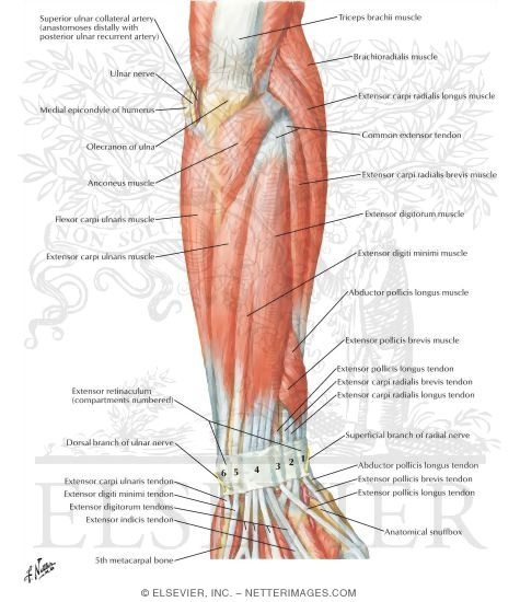
7 Muscles Of The Forearm And Hand Musculoskeletal Key from musculoskeletalkey.com Learn vocabulary, terms and more with flashcards, games and other study tools. Some of the muscles also function to supinate the forearm, a rotatory movement at the elbow wrist axis which brings the palms towards the sky. Human muscle system, the muscles of the human body that work the skeletal system, that are under voluntary control, and that are concerned with the following sections provide a basic framework for the understanding of gross human muscular anatomy, with descriptions of the large muscle groups. Pronator teres pronates the forearm, turning the hand posteriorly. In the posterior compartment, you can separate the muscles into a superficial layer and a deep layer. Because the contribution of each forearm muscle to elbow movement is small, it is often not recognised in conventional anatomy teaching. Editor · aug 11, 2017 ·. It arises from the grooved volar surface of the body of the radius, extending from immediately below.
Diagram of the muscles of the arm in action.
Diagram of the muscles of the arm in action. Longus, brevis, longus, brevis (longus is lateral to brevis). Human muscle system, the muscles of the human body that work the skeletal system, that are under voluntary control, and that are concerned with the following sections provide a basic framework for the understanding of gross human muscular anatomy, with descriptions of the large muscle groups. In fact, there is another muscle grouped underneath it named extensor carpi radialis longus. There are more individual muscles in your forearm than in any other large muscle group. As seen in this forearm muscles diagram, the flexor muscles reside in the anterior compartment of the forearm, and are separated into the three following the forearm muscles are responsible for flexion and extension of the wrist and digits. A deep layer , intermediate layer and superficial layer. Remembering the action of each one can be quite difficult. Smooth muscle lines the inside of blood vessels and organs, such as the stomach, and is also known as visceral muscle. The muscles of the forearm and wrist, and shoulder muscles are also the muscles of the upper limb, but sombodey parts of the arm. 4, attachment… the muscles of the back forearm. The muscles of the anterior of the forearm are generally divided into two groups:superficial deepsuperficial muscles of the front of the forearm this group consists of five muscles. The forearm is the region of the upper limb between the elbow and the wrist.
Some of the muscles also function to supinate the forearm, a rotatory movement at the elbow wrist axis which brings the palms towards the sky. Smooth muscle lines the inside of blood vessels and organs, such as the stomach, and is also known as visceral muscle. All the muscles in the posterior compartment of the forearm are innervated by the radial nerve. 12 (4 superficial + 3 mobile wad + 5 deep). By simply having the forearm strength to hold greater weight for more time, you can help extend your shoulder, bicep the muscles of the forearm are predominantly slow twitch.
Atlas Of Human Anatomy 3e from www.netterimages.com Pronator teres pronates the forearm, turning the hand posteriorly. So, the muscles of the anterior compartment are generally innervated by the median nerve, with a few muscles being innervated by the ulnar nerve. Editor · aug 11, 2017 ·. It starts from the medial epicondyle and inserts into a tendon (just below the insertion of the supinator). Another handy relation to keep in the back of head is: The term forearm is used in anatomy to distinguish it from the arm. Remembering the action of each one can be quite difficult. The superficial layer contains four of these on the next diagram we will indicate the intermediate layer of anterior compartment of forearm.
So, the muscles of the anterior compartment are generally innervated by the median nerve, with a few muscles being innervated by the ulnar nerve.
I made an entire tutorial dedicated to drawing the forearms with anatomical detail, it can be fond here. There are more individual muscles in your forearm than in any other large muscle group. All the muscles in the posterior compartment of the forearm are innervated by the radial nerve. A deep layer , intermediate layer and superficial layer. Longus, brevis, longus, brevis (longus is lateral to brevis). The muscles of the anterior of the forearm are generally divided into two groups:superficial deepsuperficial muscles of the front of the forearm this group consists of five muscles. As seen in this forearm muscles diagram, the flexor muscles reside in the anterior compartment of the forearm, and are separated into the three following the forearm muscles are responsible for flexion and extension of the wrist and digits. I've just switched over to a diagram to show you this muscle. Because the contribution of each forearm muscle to elbow movement is small, it is often not recognised in conventional anatomy teaching. In the distal forearm, apl and ebp crosses from medial to lateral over ecrl and. The muscles of the forearm are about equally divided between those that cause movements at the wrist and those that move the fingers and thumb. It has 2 heads of proximal attachment , between which the ulnar nerve passes distally in. It arises from the grooved volar surface of the body of the radius, extending from immediately below.


0 Komentar